
Lefort fractures, named after the French surgeon René Lefort, encompass a spectrum of severe facial injuries resulting from high-impact trauma. These fractures, classified into three primary types, necessitate a thorough understanding due to their complex anatomical involvement and varied clinical presentations.
An Elaborate Insight into Lefort Fractures and Midface Anatomy
An Elaborate Insight into Lefort Fractures and Midface Anatomy
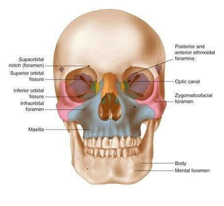
The midface region encompasses a complex network of intricately interconnected bones and delicate structures essential for facial aesthetics, sensory functions, and structural support. Comprising the maxilla, nasal bones, zygomatic bones, and the orbital floor, this intricate assembly plays a pivotal role in facial form, function, and overall harmony.
Understanding Lefort Fractures and Their Anatomical Implications
René Lefort's classification system introduced a fundamental framework for understanding midface fractures by categorizing them into three primary types, each intricately intertwined with specific anatomical patterns and distinctive clinical presentations.
Exploration of Midface Components:
Exploration of Midface Components:
Maxilla: Forming the bulk of the upper jaw, the maxilla is a central component of facial structure, housing the teeth and providing foundational support to adjacent structures. Fractures involving the maxilla can significantly impact dental occlusion and facial aesthetics | Zygomatic Bones: Often referred to as cheekbones, the zygomatic bones contribute to facial contours and the integrity of the orbital region. Fractures here can cause cosmetic deformities and compromise the stability of the orbit. |
| Nasal Bones: The paired nasal bones contribute to the nasal bridge's form and structure. Fractures in this area can lead to visible deformities, functional impairments, and nasal obstruction. | Orbital Floor: The orbital floor forms the base of the eye socket. Fractures affecting this delicate structure can result in orbital blowout injuries, causing double vision, enophthalmos (sunken eye), or restricted eye movement. |
A Comprehensive Examination of Lefort Fracture Types
A Comprehensive Examination of Lefort Fracture Types
Lefort I Fracture:
Lefort I Fracture:
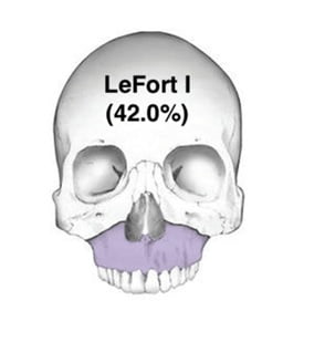
- Description: This fracture type involves a horizontal separation of the maxilla from the skull base, occurring above the level of the maxillary teeth and extending through the maxillary sinuses.
- Mechanism of Injury: Lefort I fractures typically result from direct blows to the midface region, such as those encountered in motor vehicle accidents or physical assaults.
- Clinical Characteristics: Patients may exhibit mobility of the upper jaw, anterior dental malocclusion, nasal bleeding, and subcutaneous emphysema due to communication with the maxillary sinus.
Lefort II Fracture:
Lefort II Fracture:
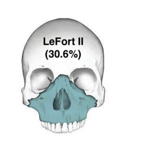
- Description: A pyramidal fracture line extending from the nasal bridge, involving the lacrimal bones, maxilla, and orbital floor, causing detachment of the entire maxilla and nasal complex.
- Etiology and Manifestations: High-energy impacts, like those occurring in falls from heights or sports-related injuries, lead to Lefort II fractures. Observable signs include periorbital ecchymosis, subconjunctival hemorrhage, and a characteristic "dish-faced" deformity.
Lefort III Fracture:
Lefort III Fracture:
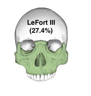
- Description: The most severe type, characterized by a transverse fracture line across the nasal bones, ethmoid bones, orbits, and zygomatic arches, completely disconnecting the facial skeleton from the skull base.
- Cause and Clinical Presentation: Lefort III fractures usually result from considerable force transmitted to the midface, causing profound facial swelling, extensive subconjunctival hemorrhage, telecanthus, and CSF leakage.
Diagnostic Approaches and Imaging Modalities:
Diagnostic Approaches and Imaging Modalities:
- Clinical Evaluation: Initial assessments involve a thorough medical history, physical examination, and evaluation for associated injuries, focusing on signs such as facial asymmetry, bruising, and malocclusion.
- Imaging Techniques: Computed tomography (CT) scans remain the gold standard for diagnosing Lefort fractures. These scans offer detailed views of fracture lines, bony displacement, and their impact on adjacent structures.
Treatment Strategies for Lefort Fractures:
Treatment Strategies for Lefort Fractures:
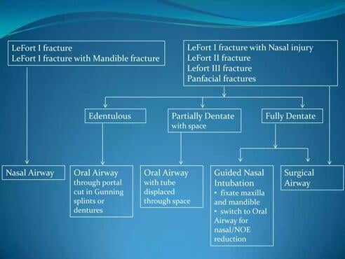
- Initial Management: Immediate stabilization and airway management are crucial in severely injured patients, often involving securing the airway and addressing any associated life-threatening injuries.
- Surgical Intervention: Definitive treatment frequently involves open reduction and internal fixation (ORIF) techniques, aiming to restore facial anatomy and function. Surgical options include the use of titanium plates, screws, or wires for stabilization and reconstruction.
Complications, Long-term Implications, and Prognosis:
Complications, Long-term Implications, and Prognosis:
- Initial Management: Immediate stabilization and airway management are crucial in severely injured patients, often involving securing the airway and addressing any associated life-threatening injuries.
- Surgical Intervention: Definitive treatment frequently involves open reduction and internal fixation (ORIF) techniques, aiming to restore facial anatomy and function. Surgical options include the use of titanium plates, screws, or wires for stabilization and reconstruction.
Conclusion:
Conclusion:
Lefort fractures represent intricate injuries that require a multidisciplinary approach for optimal management. The detailed understanding of each fracture type, their clinical presentations, diagnostic methodologies, and treatment modalities is imperative for healthcare professionals involved in trauma care and facial reconstruction.
In conclusion, the complexity and severity of Lefort fractures underscore the necessity for specialized expertise and collaborative efforts among trauma surgeons, maxillofacial specialists, radiologists, rehabilitation teams, and institutions like Malligai Dental to provide comprehensive care and achieve favorable outcomes for affected individuals. The integration of resources and expertise from facilities such as Malligai Dental is crucial in the holistic treatment and recovery journey of patients with Lefort fractures.